Add to Likebox 38220582 - BANGKOKThailand-march31 2015The doctor and staff are treating. Atrioventricular valves control blood flow between your hearts upper and lower chambers.
 A Beginner S Guide To Heart Anatomy Heart Surgery Information
Understanding heart valves anatomy is important in grasping the overall function of the heart.
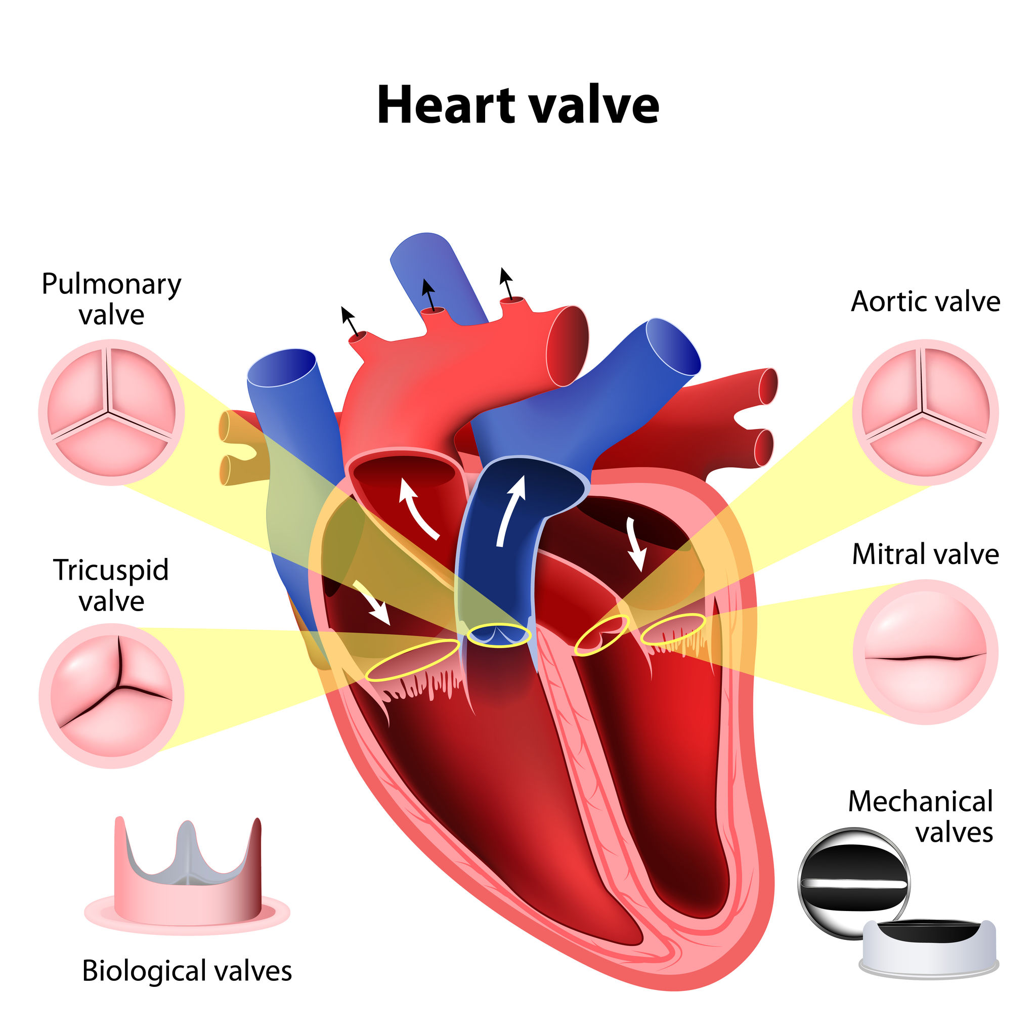
Heart valves anatomy. Dotted line marks the limit of atrial myocardium around the mitral orifice. Four valves maintain the unidirectional flow of blood through the heart. Opens to allow blood to be pumped from the right ventricle to the lungs through the pulmonary artery where it will receive oxygen.
Watch a heart valve anatomy animation. Below are the four valves of the heart. It is best to list the four valves in order of the series that blood travels through the heart.
They are composed of connective tissue and endocardium the inner layer of the heart. What are heart valves. The valves of the heart are structures which ensure blood flows in only one direction.
Separates the right ventricle from the pulmonary artery. Valves are actually flaps leaflets that act as one-way inlets for blood coming into a ventricle and one-way outlets for blood leaving a ventricle. Learn more about the future of symptomatic severe aortic stenosis management.
The tricuspid valve and. B This dissection of the heart viewed from the anterior aspect. The heart in anatomical orientation shows the spatial relations of the four cardiac valves.
97132805 - heart valves anatomy. The heart consists of four chambers two atria upper chambers and two ventricles lower chambers. Annons The role of TAVI in sAS treatment and the evidence supporting its expanding indication.
Prevents the backflow of blood as it is pumped from the left atrium to the left ventricle. The left heart valves are close together whereas the right heart valves are separated by myocardium. The valves are located between the atria and ventricles and in the two arteries that empty blood from the ventricles.
Semilunar valves control blood flow out of your heart. The heart is one of the most important organs in the body. Heart valves are flap-like structures that allow blood to flow in one direction.
The heart has two kinds of valves atrioventricular and semilunar valves. Venous blood returning from the body drains into the right atrium via the SVC IVC and coronary sinus the right atrium pumps blood through the tricuspid valve into the right ventricle. Learn more about the future of symptomatic severe aortic stenosis management.
There are four valves of the heart which are divided into two categories. Introduction to the Anatomy of the Heart Valves. These valves are actual flaps that are located on each end of the two ventricles lower chambers of the heart.
Afterwards they close during the other half of the heartbeat to ensure your blood from going backwards. The point of attachment for the heart valves. For more anatomy content please follow us and visit our website.
Prevents the back flow of blood from the pulmonary artery to the right ventricle. The Valves of the Heart The Valves of the Heart are present in 2 pairs. The valves prevent the backward flow of blood.
Heart valves are formed from elastic connective tissue which provides the flexibility needed to open and close properly. What are heart valves. These valves open and close during the cardiac cycle to direct the flow of blood through the heart chambers and out to the rest of the body.
The valve between the left atrium and the left ventricle is called the mitral valve. The heart has four valves - one for each chamber of the heart. The four valves of the heart.
The heart has 4 chambers 2 upper chambers atria and 2 lower chambers ventricles. We are pleased to provide you with the picture named Four Heart Valve Auscultation LocationWe hope this picture Four Heart Valve Auscultation Location can help you study and research. The mitral valve and tricuspid valve are located between the atria upper heart chambers and the ventricles lower heart chambers.
It is responsible for propelling blood to every organ system including itself. There is a valve through which blood passes before leaving each chamber of the heart. Also skeleton of the heart reinforced connective tissue located within the atrioventricular septum.
Prevents the backflow of blood as it is pumped from the left ventricle to the aorta. Add to Likebox 62844044 - Heart Aortic valve and atrioventricular valves. They are composed of connective tissue and endocardium the inner layer of the heart.
Other articles have discussed at length the gross anatomy of the heart and its four chambers. Heart Valves Anatomy Made of tough thin tissues named leaflets or cusps these leaflets or cusps opens during the half heartbeat to let the blood move within. The valve between the right atrium and the right ventricle is called the tricuspid valve.
Blood passes through a valve before leaving each chamber of the heart. Annons The role of TAVI in sAS treatment and the evidence supporting its expanding indication. A A pair of atrioventricular valves bring blood to the ventricles b A pair of semilunar valves drain blood from the ventricles.
Just need a glimpse leave your valuable advice let us know and subscribe us. Includes four rings that surround the openings between the atria and ventricles and the openings to the pulmonary trunk and aorta. The valves prevent the backward flow of blood.
Our LATEST youtube film is ready to run. The valves keep blood moving through the heart in the right direction.